ECR 2012 / C-1469
Cysts and cyst like renal tumors. The importance of the Bosniak classification system
Congress:
ECR 2012
Poster Number:
C-1469
Type:
Educational Exhibit
Keywords:
Cysts, Computer Applications-Detection, diagnosis, CT, Oncology, Kidney
Authors:
L. Metaxa1, I. Tsifountoudis2, P. Sountoulides1, P. Kiryttopoulos1, F. Metaxas2, M. Michaelidou2, A. Theodosiou1, I. Kalaitzoglou2; 1Veria/GR, 2Thessaloniki/GR
DOI:
10.1594/ecr2012/C-1469
 unenhanced and (B) contrast-enhanced CT scans show simple cyst in the left kidney (no enhancement)")
Fig. 2:
2. Bosniak I category: Transverse (A) unenhanced and (B) contrast-enhanced CT...
 unenhanced and (B,D) contrast-enhanced CT scans show several simple cysts in both kidneys (no enhancement, no septations, no calcification)")
Fig. 3:
3. Bosniak I category: Transverse (A,C) unenhanced and (B,D) contrast-enhanced...
 and transverse (C) contrast-enhanced CT scans show multiple simple cysts, in several sizes, in both kidneys(no enhancement, no septations, and no calcification). Also there are some cysts in the liver")
Fig. 4:
4. Bosniak I,II categories: Coronal (A,B) and transverse (C) contrast-enhanced...
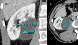
 and transverse (C) contrast-enhanced CT scans show a single, large cyst, in the right kidney with thin walls, no septa, or calcification which contains fluid with attenuation of water (2,6HU).")
Fig. 5:
5. Bosniak I category : Coronal (A) and transverse (C) contrast-enhanced CT...
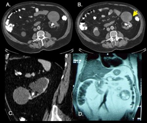
 unenhanced, (B) contrast-enhanced, Coronal (C) and , Sagittal (D) CT scans show a large cyst in the upper pole of right kidney that contains fluid with attenuation of water (9,6HU), with thin walls and hairline-thin calcified septa(yellow arrow). There is no measurable enhancement within the mass. There is another simple cyst (Bosniak I) in the lower pole of the same kidney.")
Fig. 6:
6. Bosniak II category: Transverse (A) unenhanced, (B) contrast-enhanced,...
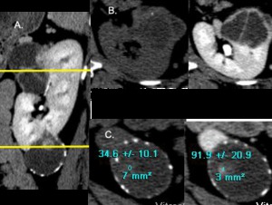
 unenhanced and (B) contrast-enhanced CT scans show a small, dense (29,6HU) mass protruding from the outermargin of the right kidney. The lesion is homogeneous and smoothly marginated and did not enhance following intravenous administration of contrast material (31HU after IV contrast media). A follow-up scan obtained 6months and a year later showed no change.")
Fig. 7:
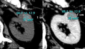
7. Bosniak IIF category: Hyperdense benign cyst; category IIF...
 unenhanced and (B) contrast-enhanced CT scans show a small hyperdense (60HU), with specific margins lesion in the parenchyma of the left kidney that lightly enhanced (less than 10HU). A follow up scan after 6, 12 and 24 months showed no important changes in size and density.")
Fig. 8:
8. Bosniak IIF category: Hyperdense benign cyst; category IIF lesion....
 unenhanced and (B) contrast-enhanced CT scans in an 82-years-old man, with chronic renal failure because of kidney and ureter stones and hydronephrosis, show an exophytic mass in the lower pole of the left kidney (C) with heterogeneity (yellow arrow), grossly thickened and irregular wall with no obvious septaes. There was a severe skepticism if the lesion was a complicated (hemorrhagic) cyst (Bosniak IIF) or a suspicious renal cystic mass (Bosniak III) and therefore had a nephrectomy. Because of the age of the patient and the medical history at first he was treated conservatively. 6-months later he had a follow up CT with no changes of the findings but a MRI scan; Coronal T2-weighted MR image (D) showed a thickened wall and multiple thickened and slightly nodular septae within the mass. The patient underwent nephrectomy and a benign complex chronic hemorrhagic renal cyst was diagnosed.")
Fig. 9:
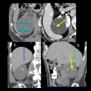
9. Bosniak III category: Transverse (A) unenhanced and (B) contrast-enhanced CT...
 cystic lesion in right kidney.Transverse (A,B,C) contrast-enhanced CT scan shows a large mostly fluid-filled mass with multiple, enhancing septa throughout. Some areas of confluence of septa, indicating solid elements, are seen.")
Fig. 10:
10. Bosniak III category: A 67-year-old man with a large (12,2x8cm) cystic...
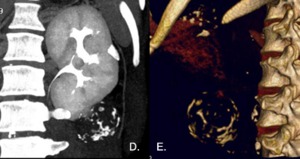
: Bosniak III-IV categories cystic lesions in a 41-years old woman with no medical history.
Coronal contrast-enhanced CT scan (A) shows two cystic lesions in the upper and lower pole of the left kidney. Transverse (B) unenhanced and contrast-enhanced CT scans at the upper pole level show a multicystic lesion with multiple septa of varying thickness. The lesion is considered to be Bosniak III category. Transverse (C) unenhanced and contrast-enhanced CT scans at the lower pole level show a single cystic lesion with thick mural calcification (Fig.11,A,C 12.D, E), and a highly enhanced nodule (34->92HU) adjacent to the wall. The lesion placed in Bosniak IV category. The patient underwent nephrectomy and a low grade clear cell carcinoma was diagnosed in both lesions on histological evaluation.")
Fig. 11:
Fig. 11-12 (same patient): Bosniak III-IV categories cystic lesions in a...
: Bosniak III-IV categories cystic lesions in a 41-years old woman with no medical history. Coronal contrast-enhanced CT scan (A) shows two cystic lesions in the upper and lower pole of the left kidney. Transverse (B) unenhanced and contrast-enhanced CT scans at the upper pole level show a multicystic lesion with multiple septa of varying thickness. The lesion is considered to be Bosniak III category. Transverse (C) unenhanced and contrast-enhanced CT scans at the lower pole level show a single cystic lesion with thick mural calcification (Fig.11,A,C 12.D, E), and a highly enhanced nodule (34->92HU) adjacent to the wall. The lesion placed in Bosniak IV category. The patient underwent nephrectomy and a low grade clear cell carcinoma was diagnosed in both lesions on histological evaluation.")
Fig. 12:
Fig. 11-12: Fig. 11-12 (same patient): Bosniak III-IV categories cystic lesions...
 reveals a large mass at the lower pole of the right kidney with septations and heterogeneity in different parts (considered to be hemorrhagic substances).
Transverse (B, D) contrast-enhanced CT scans show a cystic mass in the lower pole of the right kidney, with septations (yellow arrow) and a focal thickened (9mm) enhanced (55HU after IV contrast media)wall (C,D). No enhancement of the central portion of the mass was noted (the fluid in the central portion of the mass measures 15HU)")
Fig. 13:
13. Bosniak IV category: Cystic renal neoplasm in a 61-years old man with no...
 contrast-enhanced CT scan: A section taken through the lesion reveals a round, smooth cystic lesion with a solid enhancing nodule adjacent to the wall.")
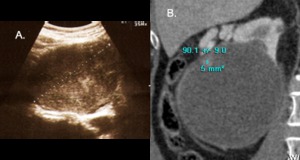
Fig. 14:
14. Bosniak IV category cystic lesion in an 80-years old man with benign...
 unenhanced and (B) contrast-enhanced CT scans reveal a round, smooth cystic lesion with some irregularity and wall thickened on its outer surface in the right kidney.")
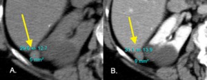
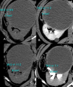
Fig. 15:
15. Same patient as Fig. 14.
Transverse (A) unenhanced and (B)...
 unenhanced and (B,D) contrast-enhanced CT scans at different levels of the cystic lesion show no enhancementof the lesion, except in the nodules at the periphery which had significant enhancement (40HU : 44,9->79,2HU and 36->78,9HU). A right nephrectomy was performed and a cystic papillary RCC filled with bloody debris was found. Viable nodules was found in the wall of the lesion. The nature of the debris apparently created the “solid” appearance on the sonogram.")
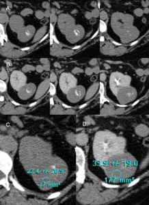
Fig. 16:
16. Same patient as Fig. 14. Transverse (A,C) unenhanced and (B,D)...
 unenhanced and (B,D) contrast-enhanced CT scans show a lesion in the upper pole of the left kidneywith high density (C)(23,4HU), mild enhancement of the central portion of the mass (D) (~10HU) and thick calcification insite the lesion. The lesion considered to be more solid rather than cystic and placed in Bosniak IV category.")
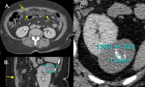
Fig. 17:
17. Bosniak IV category cystic lesion in a 60- years old woman with atypical...
 (yellow arrow) (peritoneal carcinomatosis) (A,B). A metastatic papillary renal cell carcinoma was diagnosed on histological evaluation.")
Fig. 18:
18. Same patient as Fig. 17.
Moreover enhanced nodularity along the greater...